
Listen to this Post
Introduction:
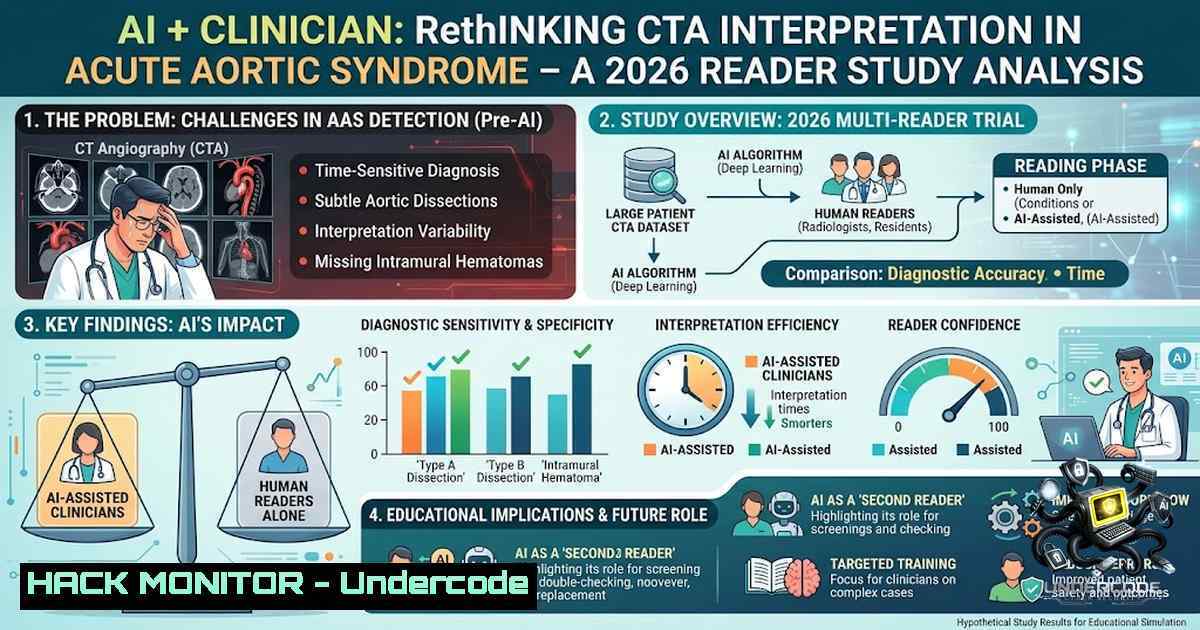
The integration of artificial intelligence into diagnostic radiology is rapidly shifting from “man versus machine” to “man and machine,” yet the metrics for success remain under scrutiny. A new multicenter reader study published in Scientific Reports on 24 August 2026 investigates this dynamic specifically for acute aortic syndrome (AAS), a life-threatening condition where every minute and every millimeter on a CTA scan counts. The study’s findings reveal a critical trade-off—higher specificity but slightly lower sensitivity—that challenges the industry to look beyond aggregate accuracy and focus on the clinical cost of missed diagnoses.
Learning Objectives & Secrets:
- Objective 1: Improve Diagnostic Specificity in Emergency Radiology – Learn how AI assistance can dramatically reduce false positives, elevating specificity from 93.80% to 98.72%. The secret tip is to calibrate the AI algorithm using a high-prevalence training set to reduce over-calling, but ensure your PACS integration highlights “low-confidence” predictions for human review.
- Objective 2: Balance Sensitivity Against Clinical Risk – Understand why a drop from 95.60% to 93.73% in sensitivity is not just a statistic but a potential liability. The secret tip is to implement a “double-read” protocol where the AI flags scans it is uncertain about, forcing a manual over-read by a second specialist to catch the missed cases without relying solely on accuracy benchmarks.
- Objective 3: Achieve Higher Inter-Reader Agreement – With the kappa score rising from 0.849 to 0.924, AI standardizes interpretation across specialties. The secret tip is to use AI-generated heatmaps and structured reports to train junior clinicians, effectively using the model as an educational overlay to reduce variance in measuring aortic diameters and intimal flaps.
You Should Know:
- Deploying AI-Assisted Workflow for CTA Interpretation in Acute Aortic Syndrome
Incorporating an AI model into a clinical workflow for AAS detection requires more than just running a Python script; it demands integration with DICOM viewers and PACS. The core objective is to automate the detection of aortic dissection, intramural hematoma, and penetrating atherosclerotic ulcer by segmenting the aorta and identifying true and false lumens.
Step‑by‑step guide explaining what this does and how to use it:
– Step 1: Data Preparation and Preprocessing – Ensure your CTA DICOMs are de-identified and converted to NIfTI format using dcm2niix. Use Linux commands to batch process: for f in .dcm; do dcm2niix -o ./output -z y $f; done. This standardizes input for the AI model.
– Step 2: Model Inference using MONAI – Deploy a pre-trained segmentation model (e.g., based on a 3D U-1et). Run inference via command line: python predict.py --input ./output/ --model aorta_net.pt --output ./results/. The model outputs probability maps for dissection presence.
– Step 3: Extracting Features for Clinical Review – Generate measurements of the maximal aortic diameter and the position of the intimal flap. Use a script to read the segmentation masks and output a CSV: python extract_metrics.py --mask ./results/mask.nii.gz --original ./output/scan.nii.gz. Export to a structured report that populates the RIS.
– Step 4: Integration with PACS – Convert the AI overlay to a DICOM secondary capture for viewing. Use `dcmtk` tools: dcmconv ai_overlay.dcm output.dcm. This ensures the radiologist sees the AI annotation directly on their diagnostic monitor alongside the native scan.
– Step 5: Auditing False Negatives – Implement a feedback loop. Store misclassified cases and run a script to flag them: grep "False_Negative" ./logs/prediction.log | mail -s "AAS Misses" [email protected]. This allows continuous monitoring of the sensitivity drop observed in the study.
- Strengthening Specificity through Threshold Tuning (ROC Curve Analysis)
The significant increase in specificity from 93.80% to 98.72% suggests the AI model used a higher confidence threshold for positive calls. While this reduces false alarms, it increases the risk of missing subtle signs. The solution is dynamic thresholding based on clinical risk factors.
Step‑by‑step guide explaining what this does and how to use it:
– Step 1: Generate ROC Curves – Using the model’s softmax probabilities, plot the ROC curves in Python (matplotlib). Calculate the optimal Youden index, but separate this for high-risk patients (e.g., Marfan syndrome) vs. low-risk.
– Step 2: Stratify Thresholds – On your inference server, set environment variables for thresholds: `export AI_THRESHOLD_HIGH=0.75` and export AI_THRESHOLD_LOW=0.45. This ensures sensitivity is not sacrificed in high-risk demographics.
– Step 3: Simulate the Reader Study – Re-run the study data using your own algorithm. Use `sklearn.metrics` to calculate sensitivity and specificity: from sklearn.metrics import accuracy_score; accuracy_score(y_true, y_pred). Compare your results against the paper’s reported 96.26% accuracy.
– Step 4: Protocol Adjustment – For patients with a clinical suspicion of AAS, implement a mandatory manual override that disregards the AI’s specificity optimization, treating any positive output as a red flag.
– Step 5: Windows/PACS Configuration – If using a Windows-based PACS, create a PowerShell script to adjust the DICOM overlay opacity based on the AI confidence, making high-confidence scans display with less overlay opacity to reduce visual clutter for the clinician.
- Mitigating the Sensitivity Drop – Strategies for Dangerous Misses
A drop to 93.73% sensitivity means that for every 100 AAS cases, ~6 are potentially missed by the AI-assisted reader. To combat this, the “Second Look” mechanism must be encoded.
Step‑by‑step guide explaining what this does and how to use it:
– Step 1: Logging Low Sensitivity Scans – Configure your AI server to create a separate log for cases where the model predicts “negative” but the output probability is between 0.4 and 0.5 (just below the high-specificity threshold). Linux command: awk '{ if ($4 >= 0.4 && $4 <= 0.5) print $0 }' ai_output.csv >> borderline_cases.csv.
– Step 2: Automated Notification – Trigger an automatic email to the on-call radiologist using `sendmail` or `msmtp` on Linux: echo "Borderline AAS case 1234" | mail -s "AI Alert" [email protected]. This prompts an immediate manual re-evaluation.
– Step 3: Visual Map Overlay – Use Python’s OpenCV to generate a heatmap of the areas where the AI was uncertain, specifically highlighting the aortic arch where flow artifacts are common. Overlay this on the original CTA.
– Step 4: Retrospective Analysis – Run a weekly script to analyze the missed cases. Install `dicom` library in Python: pip install pydicom. Use it to extract metadata (patient position, contrast phase) to identify if the sensitivity drop correlates with poor bolus timing.
– Step 5: Model Retraining – Use the false negatives to augment the training dataset. On a Windows machine with CUDA, run python retrain.py --augment false_negatives/ --epochs 10 --save model_v2.pt. This closes the loop on safety.
4. Security and API Hardening for AI Integration
Medical AI models are high-value targets. Deploying a model that can be accessed via REST API for multiple readers requires stringent security. The API endpoint likely processes DICOM files, making it susceptible to malicious payloads if not secured.
Step‑by‑step guide explaining what this does and how to use it:
– Step 1: Deploy with TLS 1.3 – Use Nginx as a reverse proxy. Configuration: `ssl_protocols TLSv1.3;` and ssl_ciphers HIGH:!aNULL:!MD5;. This encrypts data in transit.
– Step 2: API Rate Limiting – Use `nginx` or `cloudflare` to limit requests to 10 per minute per IP to prevent denial-of-service attacks that could clog the ER’s diagnostic pipeline.
– Step 3: Input Validation – Sanitize DICOM headers. Linux command to strip metadata: dcmdump -M -o sanitized.dcm original.dcm. This prevents command injection via DICOM tags.
– Step 4: Token-Based Authentication – Integrate OAuth2.0 for all API calls. Generate a JWT token: python -c "import jwt; print(jwt.encode({'user':'clinician'}, 'secret', algorithm='HS256'))". Verify tokens at the endpoint before inference begins.
– Step 5: Logging and Auditing – Enable audit logs to track who accessed which scan and when. Use Linux `auditd` to watch the inference folders: auditctl -w /var/ai/models/ -p rwx -k ai_models. This provides a chain of custody for medicolegal purposes.
- Windows and Linux Tool Configurations for the “AI + Clinician” Model
To replicate this study’s environment, one must set up a robust computing environment. Here are configurations for both operating systems to ensure the AI model runs efficiently and securely.
Step‑by‑step guide explaining what this does and how to use it:
– Linux Setup (Ubuntu 22.04) – Install CUDA 12.1 and PyTorch: `sudo apt install nvidia-cuda-toolkit` and pip install torch torchvision torchaudio --index-url https://download.pytorch.org/whl/cu121`. Verify with `nvidia-smi` to ensure the GPU is visible.docker pull projectmonai/monai:latest
- Windows Setup (Server 2022) – Enable Hyper-V and install WSL2 for Linux containers. Install Docker Desktop. Pull a MONAI Docker image:. Run the container:docker run –gpus all -p 8000:8000 monai_server.sudo docker run –1ame orthanc -p 4242:4242 -p 8042:8042 jodogne/orthanc
- Network Configuration – Ensure low latency for large CTA files. On Windows, use `netsh interface tcp set global autotuninglevel=normal` to optimize TCP/IP for large data transfers.
- DICOM Storage – Setup an Orthanc PACS server on Linux:. Configure it to store studies in/mnt/storage.sudo systemctl restart ai_inference.service`.
- CI/CD for Model Updates – Use Jenkins on Linux to auto-pull new model weights from a private GitHub repo and restart the inference service without manual intervention:
What Undercode Say:
- Key Takeaway 1: The “AI + Clinician” model is not unconditionally superior. While it significantly boosts specificity and agreement, it compromises sensitivity, meaning a 100% safety net is a myth. The clinical objective must shift to minimizing “misses” rather than maximizing “hits.”
- Key Takeaway 2: The study underscores that regulatory approval cannot be based on accuracy alone. The medical community must demand subpopulation analysis from AI vendors—specifically, which cases (e.g., intimal tears versus dissection flaps) are systematically misclassified.
Analysis: This study is a reality check for the hype surrounding AI in emergency cardiac care. The 2.47% decrease in sensitivity could translate to missed patients in a busy emergency department, leading to delayed surgery and increased mortality. While the increase in specificity (nearly 5%) reduces unnecessary follow-ups and health system costs, it introduces a trade-off that must be managed by strict workflow overrides. The future will likely see AI as a screening tool that prioritizes sensitivity (finds everything) and uses specificity to triage, rather than the current model which attempts to replicate human validation. Furthermore, the improvement in inter-reader agreement (κ to 0.924) is a significant advancement, suggesting that AI can reduce human cognitive bias and variability, making it an exceptional educational tool for residents. The security measures in deploying such models must be top-tier, as any breach or adversarial attack could manipulate specificity or sensitivity, potentially causing mass misdiagnoses. The “One Study” format of this brief reminds us that in AI, slow and cautious validation (like this multicenter reader study) remains more valuable than flashy benchmarks.
Prediction:
- +1 This study will catalyze the development of regulatory frameworks requiring AI vendors to provide “Confidence Interval” disclosures on their outputs, forcing radiology departments to link AI scores with pre-test probability algorithms.
- -1 There will be a rise in malpractice lawsuits where AI-assisted clinicians cite “AI accuracy” as a defense while missing cases, leading to a new legal standard where the “sensitivity drop” becomes a central argument against total AI reliance in acute care.
- +1 The data from this study will push hardware vendors to embed neural processing units (NPUs) directly into CT scanners, allowing for real-time “AI Second Look” during the acquisition phase, potentially recapturing the lost sensitivity by allowing the technologist to adjust the scan in real-time before the patient leaves the table.
- -1 The reliance on inter-reader agreement (κ 0.924) may inadvertently discourage clinical disagreement, leading to “groupthink” where the AI’s interpretation overrules a clinician’s suspicion, delaying the diagnosis of rarer aortic variants not covered in the training set.
- +1 Expect the release of open-source challenge datasets, similar to ImageNet, specifically for AAS, to allow developers to explicitly train for “High Sensitivity” models, decoupling the two variables to create an ensemble that flags “Actionable” vs “Surveillance” cases.
▶️ Related Video (76% Match):
🎯Let’s Practice For Free:
🎓 Live Courses & Certifications:
Join Undercode Academy for Verified Certifications
🚀 Request a Custom Project:
Secure, high-velocity infrastructure and disruptive technological engineering. Contact our engineering team for high-tier development and proprietary systems:
[email protected]
💎 Smart Architecture | 🛡️ Secure by Design | ⭐ Trusted by Thousands
IT/Security Reporter URL:
Reported By: https://lnkd.in/p/eCd78Xq8 – Hackers Feeds
Extra Hub: Undercode MoN
Basic Verification: Pass ✅


