Listen to this Post
Introduction:
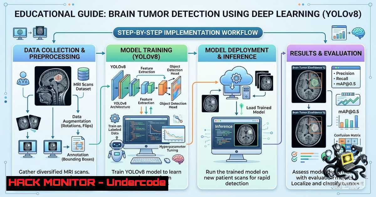
Brain tumor detection remains one of the most critical challenges in medical imaging, where early and accurate diagnosis directly impacts patient survival rates. Traditional manual analysis of MRI, CT, and PET scans is time-consuming, subjective, and prone to human error. The emergence of deep learning object detection architectures like YOLOv8 (You Only Look Once version 8) has revolutionized this space by enabling real-time, automated tumor localization with remarkable accuracy. This article presents a comprehensive technical walkthrough of implementing YOLOv8 for brain tumor detection, based on published research achieving 96.4% accuracy, covering everything from dataset preparation to production deployment in clinical environments.
Learning Objectives:
- Understand the YOLOv8 architecture and its application to medical imaging for brain tumor detection
- Learn to prepare, preprocess, and annotate medical imaging datasets for object detection training
- Implement end-to-end training, validation, and inference pipelines using Ultralytics YOLOv8
- Deploy YOLOv8 models as REST APIs with Docker containerization for clinical integration
- Apply performance optimization techniques including GPU acceleration and model quantization
1. Understanding YOLOv8 Architecture for Medical Imaging
YOLOv8 represents the latest evolution in the YOLO family of real-time object detectors, featuring a redesigned backbone, neck, and detection head that collectively deliver superior speed-accuracy trade-offs. For brain tumor detection, the architecture processes medical images by dividing them into a grid and simultaneously predicting bounding boxes and class probabilities for each grid cell. The model’s three primary components work in concert: the backbone extracts hierarchical features from input images, the neck aggregates these features across scales, and the detection head produces final predictions with class probabilities and bounding box coordinates.
What makes YOLOv8 particularly suitable for medical imaging is its anchor-free detection approach, which eliminates the need for manual anchor box design and adapts better to the irregular shapes and varying sizes of tumors. The model also introduces a new loss function that improves convergence and detection accuracy, crucial when dealing with subtle abnormalities in brain scans. The research paper highlights how YOLOv8’s real-time detection capabilities enable more efficient diagnosis and treatment planning for patients with brain tumors.
Step-by-Step Implementation:
 Install Ultralytics YOLOv8 and dependencies
pip install ultralytics opencv-python matplotlib numpy pandas

Verify installation
python -c "from ultralytics import YOLO; print('YOLOv8 installed successfully')"

Download a pre-trained YOLOv8 model weights
wget https://github.com/ultralytics/assets/releases/download/v0.0.0/yolov8n.pt
- Dataset Preparation and Annotation for Brain Tumor Detection
The quality of your dataset directly determines model performance. For brain tumor detection, medical images typically come from MRI, CT, or PET scans. The dataset must include annotated images where tumor regions are marked with bounding boxes and classified into categories such as Glioma, Meningioma, Pituitary Tumor, or No Tumor.
Data Collection and Organization:
Organize your dataset following the YOLO format requirements:
dataset/
├── train/
│ ├── images/
│ │ ├── img_001.jpg
│ │ └── ...
│ └── labels/
│ ├── img_001.txt
│ └── ...
├── val/
│ ├── images/
│ └── labels/
└── data.yaml
Annotation Format:
Each label file contains one line per object with the format: class_id x_center y_center width height, where coordinates are normalized to [0, 1].
 Example: Converting bounding box coordinates to YOLO format
def convert_to_yolo_format(bbox, image_width, image_height):
"""
Convert COCO-style bounding box [x_min, y_min, width, height]
to YOLO format [class_id, x_center, y_center, width, height]
"""
x_min, y_min, width, height = bbox
x_center = (x_min + width / 2) / image_width
y_center = (y_min + height / 2) / image_height
norm_width = width / image_width
norm_height = height / image_height
return [x_center, y_center, norm_width, norm_height]
Creating the data.yaml Configuration:
 data.yaml
path: ../dataset  dataset root directory
train: train/images  train images path
val: val/images  validation images path

nc: 4  number of classes
names: ['Glioma', 'Meningioma', 'Pituitary', 'No_Tumor']  class names
3. Preprocessing Techniques for Improved Detection Reliability
Medical images present unique challenges including noise, intensity variations, and artifacts that can degrade model performance. The research emphasizes preprocessing techniques to improve detection reliability.
Image Preprocessing Pipeline:
import cv2
import numpy as np
from skimage import exposure

def preprocess_medical_image(image_path):
"""
Complete preprocessing pipeline for brain MRI/CT images
"""
 Read image
img = cv2.imread(image_path, cv2.IMREAD_GRAYSCALE)

<ol>
<li>Noise reduction using Gaussian blur
img = cv2.GaussianBlur(img, (5, 5), 0)</p></li>
<li><p>Contrast enhancement using CLAHE (Contrast Limited Adaptive Histogram Equalization)
clahe = cv2.createCLAHE(clipLimit=2.0, tileGridSize=(8, 8))
img = clahe.apply(img)</p></li>
<li><p>Intensity normalization
img = (img - np.min(img)) / (np.max(img) - np.min(img))
img = (img  255).astype(np.uint8)</p></li>
<li><p>Convert to 3-channel for YOLOv8 (which expects RGB)
img = cv2.cvtColor(img, cv2.COLOR_GRAY2RGB)</p></li>
</ol>

<p>return img
Data Augmentation for Robustness:
from ultralytics.data.augment import Albumentations

YOLOv8 automatically applies augmentations during training
 Custom augmentation configuration can be added
augmentation_config = {
'hsv_h': 0.015,  hue augmentation
'hsv_s': 0.7,  saturation augmentation
'hsv_v': 0.4,  value augmentation
'degrees': 0.0,  rotation
'translate': 0.1,  translation
'scale': 0.5,  scaling
'shear': 0.0,  shear
'perspective': 0.0,  perspective
'flipud': 0.0,  flip up-down
'fliplr': 0.5,  flip left-right
'mosaic': 1.0,  mosaic augmentation
'mixup': 0.0,  mixup augmentation
}
4. Model Training with YOLOv8
Training the model requires careful selection of hyperparameters and monitoring of training metrics. The research paper achieved 96.4% accuracy using optimized training procedures.
Training Command:
 Single GPU training
yolo detect train data=data.yaml model=yolov8n.pt epochs=100 imgsz=640 batch=16 device=0

Multi-GPU training
yolo detect train data=data.yaml model=yolov8n.pt epochs=100 imgsz=640 batch=32 device=0,1

Training with custom hyperparameters
yolo detect train data=data.yaml model=yolov8n.pt epochs=100 imgsz=640 batch=16 \
optimizer=AdamW lr0=0.001 weight_decay=0.0005 momentum=0.937
Python Training Script:
from ultralytics import YOLO

Load a pre-trained model
model = YOLO('yolov8n.pt')  nano version for speed
 model = YOLO('yolov8m.pt')  medium for better accuracy
 model = YOLO('yolov8l.pt')  large for maximum accuracy

Train the model
results = model.train(
data='data.yaml',
epochs=100,
imgsz=640,
batch=16,
device=0,  GPU device
workers=8,
patience=50,  early stopping patience
save=True,
save_period=10,  save checkpoint every 10 epochs
val_period=1,  validate every epoch
project='brain_tumor_detection',
name='yolov8n_brain_tumor'
)

Evaluate the model
metrics = model.val()
print(f"mAP50: {metrics.box.map50:.4f}")
print(f"mAP50-95: {metrics.box.map:.4f}")
Training Monitoring with TensorBoard:
 Launch TensorBoard to monitor training
tensorboard --logdir runs/detect

Or use the built-in YOLO logging
yolo detect train data=data.yaml model=yolov8n.pt epochs=100 project=logs
5. Model Validation and Performance Metrics
Proper validation ensures the model generalizes well to unseen medical images. The research emphasizes robust evaluation metrics for medical validation.
Validation Command:
 Validate the trained model
yolo detect val data=data.yaml model=runs/detect/train/weights/best.pt

Validate with specific metrics
yolo detect val data=data.yaml model=best.pt conf=0.25 iou=0.45
Comprehensive Evaluation Script:
from ultralytics import YOLO
import numpy as np
from sklearn.metrics import confusion_matrix, classification_report

Load trained model
model = YOLO('runs/detect/train/weights/best.pt')

Run validation
results = model.val(data='data.yaml')

Extract metrics
print(f"Precision: {results.box.mp:.4f}")
print(f"Recall: {results.box.mr:.4f}")
print(f"mAP50: {results.box.map50:.4f}")
print(f"mAP50-95: {results.box.map:.4f}")

Per-class performance
for i, class_name in enumerate(['Glioma', 'Meningioma', 'Pituitary', 'No_Tumor']):
print(f"{class_name} - Precision: {results.box.p[bash]:.4f}, Recall: {results.box.r[bash]:.4f}")
Confusion Matrix Generation:
 Generate confusion matrix for detailed analysis
from ultralytics.utils.metrics import ConfusionMatrix

Create confusion matrix
conf_matrix = ConfusionMatrix(nc=4, conf=0.25)
 ... populate with predictions
6. Real-Time Inference and Deployment
The ability to perform real-time inference is crucial for clinical deployment. The research highlights fast inference performance suited for real-time use.
Inference on Single Images:
from ultralytics import YOLO
import cv2

Load the trained model
model = YOLO('runs/detect/train/weights/best.pt')

Run inference on an image
results = model('path/to/mri_scan.jpg')

Display results
for r in results:
boxes = r.boxes
for box in boxes:
x1, y1, x2, y2 = box.xyxy[bash].tolist()
conf = box.conf[bash].item()
cls = int(box.cls[bash].item())
print(f"Tumor detected at ({x1:.0f}, {y1:.0f}) - ({x2:.0f}, {y2:.0f}) with confidence {conf:.2f}")
Real-Time Video/Webcam Inference:
import cv2
from ultralytics import YOLO

model = YOLO('best.pt')

Open webcam
cap = cv2.VideoCapture(0)

while True:
ret, frame = cap.read()
if not ret:
break

Run inference
results = model(frame)

Annotate frame
annotated_frame = results[bash].plot()

Display
cv2.imshow('Brain Tumor Detection', annotated_frame)

if cv2.waitKey(1) & 0xFF == ord('q'):
break

cap.release()
cv2.destroyAllWindows()
Flask API for Clinical Integration:
from flask import Flask, request, jsonify
from ultralytics import YOLO
import cv2
import numpy as np
import base64

app = Flask(<strong>name</strong>)
model = YOLO('best.pt')

@app.route('/detect', methods=['POST'])
def detect_tumor():
 Get image from request
data = request.get_json()
image_data = base64.b64decode(data['image'])
nparr = np.frombuffer(image_data, np.uint8)
img = cv2.imdecode(nparr, cv2.IMREAD_COLOR)

Run inference
results = model(img)

Extract predictions
predictions = []
for r in results:
for box in r.boxes:
x1, y1, x2, y2 = box.xyxy[bash].tolist()
conf = box.conf[bash].item()
cls = int(box.cls[bash].item())
predictions.append({
'bbox': [x1, y1, x2, y2],
'confidence': conf,
'class': ['Glioma', 'Meningioma', 'Pituitary', 'No_Tumor'][bash]
})

return jsonify({'predictions': predictions})

if <strong>name</strong> == '<strong>main</strong>':
app.run(host='0.0.0.0', port=5000)
7. Docker Containerization for Production Deployment
Containerization ensures consistent deployment across clinical environments.
Dockerfile:
FROM python:3.9-slim

WORKDIR /app

Install system dependencies
RUN apt-get update && apt-get install -y \
libgl1-mesa-glx \
libglib2.0-0 \
&& rm -rf /var/lib/apt/lists/

Copy requirements and install Python dependencies
COPY requirements.txt .
RUN pip install --1o-cache-dir -r requirements.txt

Copy model and application
COPY best.pt .
COPY app.py .

Expose port
EXPOSE 5000

Run the application
CMD ["python", "app.py"]
requirements.txt:
ultralytics>=8.0.0
opencv-python>=4.5.0
flask>=2.0.0
numpy>=1.19.0
Build and Run:
 Build Docker image
docker build -t brain-tumor-detector .

Run container
docker run -d -p 5000:5000 --1ame brain-tumor-api brain-tumor-detector

Save for offline deployment
docker save brain-tumor-detector > brain-tumor-detector.tar
8. Model Optimization for Clinical Deployment
For production environments, model optimization is essential for low-latency inference.
Model Export to ONNX:
 Export to ONNX format
yolo export model=best.pt format=onnx imgsz=640

Export with dynamic batch size
yolo export model=best.pt format=onnx imgsz=640 dynamic=True
Model Quantization for Edge Deployment:
from ultralytics import YOLO

Load model
model = YOLO('best.pt')

Export to TensorRT for NVIDIA GPU acceleration
model.export(format='engine', device=0)

Export to TFLite for mobile/edge deployment
model.export(format='tflite', imgsz=640)

Export to OpenVINO for Intel optimization
model.export(format='openvino', imgsz=640)
Inference with Optimized Model:
 Load optimized model
model = YOLO('best.engine')  TensorRT
 model = YOLO('best_openvino_model')  OpenVINO

Run inference (significantly faster)
results = model('mri_scan.jpg')
What Undercode Say:
- Key Takeaway 1: The integration of YOLOv8 with medical imaging represents a paradigm shift in diagnostic capabilities. With 96.4% accuracy demonstrated in published research, this approach bridges the gap between cutting-edge computer vision and clinical practice, enabling radiologists to make faster, more accurate diagnoses. The real-time inference capability means that what once took hours of manual analysis can now be accomplished in milliseconds.
-
Key Takeaway 2: The technical implementation requires careful attention to medical image preprocessing, dataset quality, and model validation. The success of this approach depends not just on the algorithm but on robust data pipelines, proper annotation, and thorough validation against ground truth. The combination of YOLOv8’s architectural advantages with specialized preprocessing techniques creates a system that can reliably detect tumors across diverse imaging modalities and patient populations.
Analysis: The research demonstrates that deep learning, specifically YOLOv8, can achieve clinically relevant accuracy for brain tumor detection. The 96.4% accuracy rate suggests that such systems are approaching the diagnostic capabilities of experienced radiologists, though they should be viewed as assistive tools rather than replacements. The real-time performance opens possibilities for intraoperative imaging, emergency department screening, and telemedicine applications in underserved regions. However, challenges remain in handling the diversity of tumor presentations, addressing class imbalance in datasets, and ensuring model robustness across different MRI machines and protocols. The translational implications highlighted in the research suggest that these technologies are moving from research labs to clinical practice, with the potential to significantly improve patient outcomes through earlier detection and more precise treatment planning.
Prediction:
- +1 Clinical adoption of YOLOv8-based detection systems will accelerate dramatically over the next 2-3 years, driven by the proven accuracy and real-time capabilities demonstrated in peer-reviewed research. Integration with existing PACS (Picture Archiving and Communication Systems) will become standard, enabling seamless AI-assisted diagnosis workflows.
-
+1 The combination of YOLOv8 with radiogenomics, as mentioned in the research, will unlock new possibilities for personalized medicine by correlating imaging features with genomic markers, potentially revolutionizing treatment planning and prognosis prediction.
-
-1 Regulatory challenges and the need for extensive clinical validation across diverse patient populations may slow widespread deployment. Healthcare institutions will need to navigate complex FDA/CE approval processes and establish protocols for AI-assisted diagnosis liability.
-
+1 Edge deployment on portable devices and mobile platforms will democratize access to advanced diagnostic capabilities, particularly benefiting telemedicine and healthcare delivery in resource-limited settings.
-
-1 The risk of algorithmic bias and performance degradation across different imaging equipment manufacturers and protocols remains a significant concern that requires ongoing monitoring and model retraining strategies to ensure equitable patient care.
▶️ Related Video (82% Match):
https://www.youtube.com/watch?v=1tPrUGDz4Uk
🎯Let’s Practice For Free:
🎓 Live Courses & Certifications:
Join Undercode Academy for Verified Certifications
🚀 Request a Custom Project:
Secure, high-velocity infrastructure and disruptive technological engineering. Contact our engineering team for high-tier development and proprietary systems:
[email protected]
💎 Smart Architecture | 🛡️ Secure by Design | ⭐ Trusted by Thousands
IT/Security Reporter URL:
Reported By: Kesava Rao – Hackers Feeds
Extra Hub: Undercode MoN
Basic Verification: Pass ✅


